what is best to use on a pressure ulcer
Wound and Pressure Ulcer Direction
past Sharon DeMarco, CRNP
- Introduction
- Prevention
- Assessment
- Wound Healing
- Treatment
- References
Introduction
Education of patients, families, caregivers and healthcare providers is the key to a proactive program of prevention and timely, appropriate interventions (Erwin-Toth and Stenger 2001). Wound direction involves a comprehensive care plan with consideration of all factors contributing to and affecting the wound and the patient. No single discipline can encounter all the needs of a patient with a wound. The best outcomes are generated by dedicated, well educated personnel from multiple disciplines working together for the common goal of holistic patient care (Gottrup, Nix & Bryant 2007).
Significance of the problem:
- Pressure ulcer (Pr U) incidence is associated with an increased Morbidity & Mortality – nearly 70% die within six months. (Brown 2003)
- Pr U incidence is increasing in long term care. (LTC) (Horn et al. 2004)
- Reduction of pressure ulcer prevalence in LTC is a Healthy People 2010 initiative.
- Pr U incidence has been determined to be a quality of intendance indicator for LTC facilities and compliance is regulated past the Center for Medicare and Medicaid.(CMS 2004)
- Lawsuits due to Pr Us are on the ascension. (Voss et al. 2005)
- Leg ulcers affect more individuals than Pr Us; one in four Americans over the historic period of 65 will develop a leg ulcer in their lifetime (Wound Ostomy and Continence Nurses Society [WOCN] 2002)
- Skin and wound allegations are the second leading crusade of litigation in LTC. (Chizek 2003)
Prevention
- Anatomy of normal skin
- What is a pressure ulcer?
- How do you prevent a pressure ulcer?
- What is Not a pressure ulcer?
Anatomy of Normal Skin

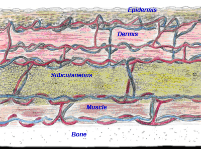
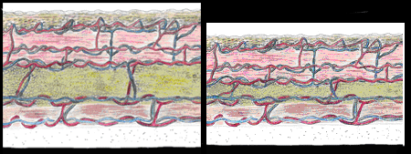
Age related pare changes (meet comparison figures below-normal on the left, aging on the correct) include thinning and atrophy of epithelial and fat layers. Additionally, collagen and elastin compress and degenerate, and dermal fibroblasts cease replicating, all resulting in thinner, drier and less elastic peel that heals more slowly.

(summit of section)
(tiptop of page)
What is a Pressure Ulcer?
Previously called decubitus or bed sore, a pressure ulcer is the result of harm caused by force per unit area over fourth dimension causing an ischemia of underlying structures. Bony prominences are the nearly common sites and causes.
There are many risk factors that contribute to the development of pressure ulcers. CMS (2004) recommends patients in LTC be assessed for run a risk on admission, weekly for the first 4 weeks and so reassessed quarterly.
At that place are many contributing factors.
Intrinsic contributing factors include:
- Malnutrition
- Dehydration
- Impaired mobility
- Chronic conditions
- Impaired sensation
- Decreased LOC
- Infection
- Advance age
- Steroid use
- Pressure ulcer present
External contributing factors include:
- Pressure level
- Friction
- Moisture
- Incontinence
- Shear
(height of department)
(top of page)
How Practise You lot Forestall a Pressure Ulcer? (WOCN 2003; AHCPR 1992)
Proper skin care is crucial and involves inspecting skin daily and an individualized bathing schedule, using warm (not hot) water and mild lather. Avoid massage over bony prominences and utilise lubricants if skin is dry out.
Managing pressure is also necessary and the following is recommended.
- Provide appropriate support surface
- Reposition every 2 hours in bed
- Off-load heels - utilise pillows or positioning boot
- Reposition every hour when in chair
- Use pillow between legs for side lying
- Practise non position directly on trochanter
- Do non employ doughnut-type devices
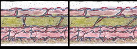
Friction and shear need to be reduced. Friction is the mechanical strength exerted when skin is dragged against a coarse surface while shear is the mechanical force caused by the interplay of gravity and friction. It exerts a forcefulness parallel to the skin resulting in angulation and stretching of blood vessels (shown below on right) within the sub-dermal tissues, causing thrombosis and cellular death. This manifests equally necrosis and undermining of the deepest layers (Pieper 2007).

To reduce friction and shear, the following is recommended:
- Use depict sheets for repositioning
- Encourage utilise of trapeze if possible
- Keep caput of bed elevated ? thirty? if tolerated
- Drag human foot of bed slightly, if condition permits
- Employ pillow or wedge to support hip for xxx? side-lying, lateral position
- Utilise lifts and transfer devices
- Rehabilitation or Restorative care if indicated
Manage Incontinence
- Timely cleansing
- Apply barrier ointment to intact skin
- If skin is carmine or denuded apply a paste
- Use advisable incontinence disposables
- Apply fecal incontinence pouch if needed
What is Not a Pressure Ulcer?
Peel tears, denuded or excoriated peel, arterial ulcers, venous stasis ulcers and diabetic/neurotrophic ulcers are Non pressure level ulcers.
Peel Tear Prevention (Ayello 2003)
- Wash with gentle cleansing products
- Use emollients on skin
- Ensure acceptable hydration/diet
- Transfer techniques to avert friction/shear
- Support dangling extremities
- Avoid use of agglutinative products on skin
Venous Ulcer Prevention (Vowden & Vowden 2006)
- Use of pinch stockings (contraindicated if ABI < 0.5)
- Elevation of effected leg higher up level of the center at rest
- Avoid use of products probable to be sensitizers (lanolin, fragrances)
- Avert trauma to legs
- Calf musculus strengthening
- Regular follow up to monitor ABI
Prevention of Limb loss in Lower extremity arterial disease (Hopf et al. 2006)
- Consistent use of protective footwear
- Avoid friction, shear or trauma to feet/legs
- Utilise emollients to keep skin pliable
- Avoid common cold, caffeine, nicotine and constrictive garments
- Planned graduated walking programme
- No apply of thermal devices
- Routine professional person human foot intendance
Prevention of neuropathic ulcers (Steed et al. 2006)
- Control diabetes
- Daily care and inspection of feet
- Wear well-plumbing fixtures protective footwear
- Avoid application of external rut
- Avoid employ of OTC meds for corns/callous
- Avoid cold, caffeine, nicotine and constrictive garments
- Routine professional foot care
(elevation of department)
(tiptop of page)
Cess (Nix 2007)
- Holistic Cess
- Wound Assessment
- Additional assessment for lower extremity wounds
Holistic Assessment
Holistic cess of a patient with a wound includes systemic factors, psychosocial factors, and local factors.
Systemic factors assess etiology, duration, and decreased oxygenation or perfusion of the wound likewise as comorbid conditions, medications, and host infection of the patient.
Psychosocial factors to address in a holistic cess include the patient'southward knowledge deficits, cultural beliefs and fiscal constraints including a lack of or insufficient health insurance. Additionally, it is necessary to assess whether the patient has dumb admission to appropriate resource and any social support – family, pregnant others or community resource.
Local factors to assess include desiccation, backlog exudates, low wound temperature, recurrent trauma (as well friction & pressure), infection, and necrosis and foreign bodies.
Wound Assessment
An assessment of the wound should be done weekly and exist used to drive handling decisions. Wound cess includes: location, grade/stage, size, base tissues, exudates, odor, edge/perimeter, pain and an evaluation for infection.
Location
Documentation of location indicating which extremity, nearest bony prominence or anatomical landmark is necessary for appropriate monitoring of wounds. (Hess 2005)
Form/Stage
Pressure ulcers are classified past stages equally defined by the National Force per unit area Ulcer Advisory Panel (NPUAP). Originally there were iv stages (I-Four) but in February 2007 these stages were revised and two more categories were added, deep tissue injury and unstageable.
Pressure Ulcer Staging
Stage I - Intact skin with non-blanchable redness of a localized surface area, usually over a bony prominence. Darkly pigmented peel may non accept visible blanching; its color may differ from the surrounding surface area.
Stage 2 - Partial thickness loss of dermis presenting as a shallow open ulcer with a red/pink wound bed, without slough. May also nowadays as an intact or open up/ruptured serum filled blister.
Phase III - Full thickness peel loss. Subcutaneous fat may be visible but bone, tendon or muscle are not exposed. Slough may be present but does non obscure the depth of tissue loss. May include undermining/tunneling.
Phase IV - Full thickness skin loss with exposed os, tendon or muscle. Slough or eschar may be present on some parts of the wound bed. Oft include undermining and tunneling.
Unstageable - Full thickness tissue loss in which the base of the ulcer is covered by slough (yellow, tan, gray, green or brown) and/or eschar (tan, chocolate-brown or blackness) in the wound bed.
(Suspected Deep) Tissue Injury - Royal or maroon localized area of discolored intact skin or claret-filled blister due to impairment of underlying soft tissue from pressure level and/or shear. The expanse may be preceded by tissue that is painful, firm, mushy, boggy, warmer or cooler equally compared to adjacent tissue. (NPUAP 2/07)
Class
At that place are a number of classification and grading systems used in wound care but the simplest method uses the terms partial thickness or full thickness
• Fractional thickness wound (PTW): damage to epidermis and/or dermis only
• Full thickness wound (FTW): damage to subcutaneous layer or deeper
Size
Measurement
- Length – from top border to the bottom edge (head to toe) at longest indicate
- Width – from border to border perpendicular to the length at widest indicate
- Depth – straight in, perpendicular to the base of operations, at deepest point
Undermining/Tunneling
- Using the "clock concept" (12 o'clock is in the direction of the patient's head and 6 o'clock is toward the feet)
- Where does information technology showtime and where does it finish (clockwise direction)
- Tunnel depth is at it's deepest point
- Location of deepest signal
Base Tissues
Assessing the advent of tissue in the wound bed is critical for determining appropriate treatment strategies and to evaluate progress toward healing. (Keast et al. 2004)
Necrosis/Eschar - Black, dark-brown or tan devitalized tissue that adheres to the wound bed or edges and may be firmer or softer than the surrounding pare.
Slough - Soft, moist avascular tissue that adheres to the wound bed in strings or thick clumps; may be white, xanthous, tan or green.
Granulation - Pink/red moist tissue comprised of new blood vessels, collagen fibers and fibroblasts. Typically the surface is shiny and moist with a granular appearance.
Epithelium - New pink and shin tissue/skin that grows in from the edges or every bit islands on the wound surface.
Exudates
Amount
- None – base and dressing dry out
- Slight – small-scale amount in eye of dressing
- Moderate – contained within the dressing
- Copious – extends beyond dressing onto clothing or bed linen
Type
- Serous – sparse, watery, clear or straw colored
- Serosanguineous – thin, pale carmine to pinkish
- Purulent – thick, opaque, tan, yellowish to light-green and may accept an offensive odor
- Consider treatment modality and frequency of dressing changes
Odor
Assess after cleansing (Garcia & Thomas 2006). Extreme malodor, peculiarly if accompanied past purulent exudates is suggestive of infection. Most wounds exercise take an odor. The type of dressing can affect aroma as well as hygiene and the presence of nonviable tissue (Keast et al. 2004).
Edge/Perimeter
- Describe wound edges (approximated, rolled, calloused)
- Describe periwound skin (indurated, erythematous, diminished, healthy)
- Describe presence of excoriation, denudement, erosion, papules, pustules or other lesions
Induration - Abnormal hardening of the tissue caused by consolidation of edema,
this may be a sign of underlying infection.
Erythema - Redness of surrounding tissue may be normal in the inflammatory phase of healing. However, if accompanied by an increase in temperature of tissue, exudates or pain may also be a sign of infection.
Maceration - Caused by excessive moisture, Tissue loses its pigmentation (appears lucid or turns white) and becomes soft and friable.
Pain
A critical aspect of local wound assessment both from the perspective of the patient and as a clinical indicator of infection. (Reddy, Keast, Fowler & Sibbald 2003) Include location, blazon/cause, rating (employ validated scale), patient description and nonverbal signs.
Evaluation of infection
Infection – Signs and Symptoms:
- Redness, warmth and induration of adjacent tissues
- Hurting or tenderness
- Dysmorphic and/or friable granulation
- Unusual odor
- Purulent exudates
- Systemic signs (fever, chills, sweats)
When to Civilisation: (Dow 2003)
- When signs of infection are present or when a clean wound fails to heal
- Always cleanse wound starting time
- Semi-quantitative swab collection is adequate
- Quantitative biopsy is "gold standard" simply expensive and invasive
(top of section)
(top of page)
Additional Assessment for Lower Extremity Wounds (WOCN 2002)
Physical Examination
- Edema –extent and persistence of pitting (1+ - 4+)
- Colour changes - dependent rubor (purple-carmine discoloration) or elevation pallor (paling of the skin when leg raised to a 60° angle for 15 -threescore seconds)
- Distal pulses –amplitude on palpation (0 – 4+)
- Neuropathy – skin changes (dryness, corking), structural abnormalities, and loss of protective sensation (10gm monofilament examination – testing x points)
Diagnostic Tests
- Ankle-Brachial index – comparing of perfusion pressures
- Pulse volume recording - perfusion volume
- Doppler waveforms – single vessel flow
- Duplex imaging – ultrasound imaging for venous illness (besides exam for DVT)
- Transcutaneous oxygen pressure (TcPO2)
(superlative of section)
(top of folio)
Wound Healing
- Phases of Wound Healing
- Optimization of Wound Environment
Phases of Wound Healing
There are three phases of wound healing - inflammation, proliferation, maturation
Inflammatory Stage
- 0 – iii days
- Hemostasis (bleeding stops)
- Inflammation (redness, swelling, warmth and pain maybe present)
- Phagocytosis (WBC's engulf bacteria and foreign droppings)
- Growth gene stimulation stimulation
Proliferation Stage
- 3 – 21 days
- Angiogenesis (new blood vessels develop)
- Collagen synthesis (protein fibers)
- Granulation formation
- Epithelialization
- Contraction
Maturation Phase
- 21 days – 2 years
- Reorganization of collagen
- Tensile strength improves (up to 80% of original)
The healing procedure varies depending on the stage of the pressure ulcer. Phase I & 2 pressure ulcers and fractional thickness wounds heal by tissue regeneration. Phase III & Iv pressure level ulcers and full thickness wounds heal by scar formation and wrinkle. Data indicate a 20% reduction in wound size over two weeks is a reliable predictive indicator of healing. (Flanagan 2003)
(superlative of section)
(top of page)
Optimization of Wound Environment
- Manage comorbid weather condition
- Adequate nutrition & hydration
- Remove nonviable tissue
- Maintain moisture residue
- Protect the wound and periwound peel
- Eliminate or minimize pain
- Cleanse
- Foreclose and manage infection
- Control odor
Manage comorbid condition
- Optimize cardiovascular and pulmonary functioning
- Support tissue oxygenation
- Maintain blood glucose control
Adequate nutrition & hydration (Harris & Frasier 2004)
- Encourage protein, calorie-dense foods and fluids, unless contraindicated
- Monitor intake, weight and skin turgor
- Assess and accost impairments in dentition and swallowing
- Assistance patients with meals if needed
- Dietary consult
Eliminate or Minimize Hurting
- Address the crusade (remove the source if external, treat the infection or medicate based on physiological stimulus)
- Pharmacological strategies –long interim drugs preferable, use breakthrough doses and prevent agin effects
- Contain psycho-social, spiritual and culturally sensitive support
- Advisable dressing selection, gentle removal and "Time out" during treatment administration
Cleanse
- Normal saline is the recommended solution
- Cavity wounds or tunnels may exist irrigated
- Apply 4-15 (psi) pressure level/forcefulness to remove droppings without harming healthy tissue
Protect Wound and Periwound Skin
- Utilise bulwark products to protect from adhesives and wet
- Change dressings at appropriate intervals to avert pooling of exudates
Prevent and Manage Infection
Disquisitional colonization tin can result in failure to heal, poor quality tissue, increased friability and increased drainage (Frank, Bayoumi & Westendorp 2005). Determining whether the wound has a bacterial imbalance (critical colonization and infection) is of primary importance to healing (Sibbald, Woo & Ayello 2006).
- Superficial increased bacterial burden – topical agent with low toxicity, non likely to cause allergy and non associated with bacterial resistance
- Surrounding pare compartment infection – topical agent, swab civilization and advisable oral antibody amanuensis (Sibbald 2003)
- Deep wound infection or osteomyelitis – parenteral antibiotics. Also consider tissue culture and boosted lab tests (Frank et al. 2005)
Removal of Nonviable Tissue (Debridement)
Removes growth medium, controls/ prevents infection, defines extent of the wound and stimulates the healing procedure.
Contraindications
-
Dry stable heel eschar
-
Ischemic wounds with dry gangrene
-
Coagulation disorders
Types of Debridement
-
Autolytic debridement
-
-
Lysis of necrotic tissue by the body's white blood cells and enzymes.
-
Leaves healthy tissue intact
-
Naturally occurring physiological process that occurs in a moist environs
-
-
Chemical Debridement - Achieved by topical awarding of enzymes:
-
-
Collagense (Santyl®)
-
Papain with urea (Accuzyme®, Ethezyme®, Gladase®)
-
Denaturing agents are likewise used: Sodium hypochlorite (Chlorpactin®, Dakin'southward) Annotation: This is a nonselective method
-
-
Mechanical Debridement
-
-
Wet to dry dressing (non recommended as it is nonselective, causes repeated trauma to the wound bed and is frequently painful)
-
Whirlpool (gamble of cross contamination and contraindicated for some wounds such every bit venous stasis)
-
Pulse lavage (requires skilled clinician, rigorous infection control precautions and may be cost prohibitive
-
-
Precipitous Debridement
-
-
Conservative, sequential removal of avascular tissue, using sterile scalpel and mouse-molar forceps
-
Check for bleeding or clotting problems
-
Pre-medicate if wound is painful
-
Avoid local anesthetic
-
-
Surgical Debridement
Maintain Moisture Residue (Rolstad & Ovington 2007)
- Dressings with a high moisture vapor manual rate will permit wet to escape and evaporate in minimally exudative wounds
- Moderate to heavily draining wounds require absorptive dressings
Control Scent
- Appropriate frequency of dressing changes
- Cleanse with each dressing change
- Debridement and antimicrobials as indicated
- Charcoal dressings
(elevation of section)
(top of page)
Treatment
- Objectives and Plan
- Palliative Wound Care
- Factors for Dressing Pick
- Production Categories
Objectives and Plan
The provider's role is to assist in the evolution of a sustainable programme designed to assist achieve mutually agreed upon goals. (Cypher & Pierce 2007) Treatment goals should be identified and can be curative or palliative. Palliative care objectives focus on symptom management and quality of life.
The objectives vary depending on the staging of the wound:
- Recently closed wound, Stage I pressure ulcer, denuded or excoriated skin - Encourage adequate perfusion and protect from further tissue damage.
- Stage II or PTW - Encourage regeneration of tissue and protect wound surface.
- Phase III/IV - Promote granulation and wrinkle (epithelialization)
Palliative Wound Intendance (Bradley 2004)
- Symptom management: elimination or reduction of hurting, control of olfactory property and exudates, treatment/prevention of infection
- Quality of life objectives: restoration of some sense of command, maintenance of function and independence, control of caregiver burden, reduction of distress for patient and family.
(pinnacle of section)
(summit of page)
Factors for Dressing Selection
- Etiology
- Exudates
- Wound history
- Odor
- Comorbid weather
- Perimeter
- Size
- Patient/caregiver needs
- Base
- Admission
Etiology - The cause of the wound directly affects dressing choices. For example:
- Arterial ulcers generally require moisture
- Neuropathic wounds ofttimes have tunnels which require packing strips
- Pressure ulcers frequently accept undermining which requires packing to fill expressionless space
- Venous insufficiency requires compression and exudate management
Wound History
- Elapsing and class of wound healing
- Previous dressings/treatment strategies
- Health intendance providers consulted for wound
- Success/challenges of previous handling
Comorbid conditions
- Diabetes – impairs wound healing, compromises perfusion and there is an increased take a chance of infection.
- Mixed (arterial and venous disease) or CHF - pinch may be contraindicated
- Obesity – increased hazard of venous hypertension, infection and dehiscence (Wilson & Clark 2003)
- Immunosupression – increases take chances of infection and impairs healing
Size
- Size and extent of tissue loss determines both the dressing size and fabric
- Wound packing needed for larger wounds
- Exposed tendons/ligaments crave moisture and protection
Base of operations
- Clean healthy granulation – keep moist
- Slough – debridement: If slight amount, keep moist to encourage autolysis, If there is a moderate corporeality, utilize a chemical or mechanical agent. For large amounts, perform series sharp debridement and may besides apply adjunct treatment with chemical or mechanical amanuensis.
- Epithelium- moist protective dressing
Exudates
- The book and type of exudates are significant determinants in selection of primary and secondary dressings
- Adequate containment of exudate is critical to manage increased bioburden , protect the periwound pare, control odor and avoid overuse of wound care products (Rolstadt & Ovington 2007)
Olfactory property
- Commonly associated with an infected wound
- Fungating lesions or wounds with high colonization due to necrotic droppings may be malodorous
- Aroma may cause considerable patient/ caregiver stress and embarrassment
Perimeter - Status of the periwound skin influences the blazon of products used and may indicate the need for additional products.
- Barriers are indicated for frail or compromised skin
- Maceration indicates need for exudate management
- Topical treatment may be required for fungal infections
Patient/caregiver needs
- Who is providing the care?
- Do they take cerebral, dexterity or visual impairments to consider?
- In what setting volition the dressings be done?
- Are didactics and/or training needed?
- Are wellness intendance resources available?
- Is the handling plan coinciding with the culture/beliefs of the patient/caregiver?
Admission
- Does the patient have access to supplies and services?
- Are there financial constraints or limitations with insurance coverage?
- Is transportation a factor in accessing intendance or supplies?
Product Categories (Sibbald 2003) (Okan et al. 2007) (Cipher 2007)
There are a neat bargain of products focused on wound management. Below is a breakup of products by their function in wound and ulcer care.
Antimicrobials (topical)
- Bacitracin®, - Broad spectrum, low price, apply daily.
- Bactroban®) - Splendid penetration, effective for MRS. Use three times daily.
- Cadexomer Iodine (Iodosorb®) – Contains microspheres that blot bacteria while slowly releasing iodine and is less toxic to granulation. Broad spectrum, including virus and fungus. Effective for up to 72 hours.
- Nanocrystalline silver (Acticoat seven®) – Releases bactericidal concentrations up to seven days. Use sterile water, not saline. May stain peel. May be toll prohitive.
- Polysporin® powder – For Gram-negative and Gram positive organisms and Pseudomonas. May apply with Santyl®. Apply daily.
- Silvery sulfadiazine (Silvadene®) cream – Wide spectrum. Cost constructive, but requires prescription. Avoid in sulfa allergy. Apply daily.
- Silver impregnated hydrofiber (Aquacel Ag®) – Highly absorbent. Silver stays in dressing, very picayune is deposited into wound base. Change when saturated
- Silvery gel (SilvaSorb®) – Wide-spectrum and low toxicity. Delivers time-released silverish for 3 days.
- Sodium hypochlorite (Chlorpactin®) – Most advisable for malodorous wounds with big amounts of slough. Twice daily for short term treatment only (less than ten days).
Alginates
- Derived from seaweed
- Highly absorbent and biodegradable
- Hemostatic backdrop
- Conforms to wound shape
- Maintains moist environment
- Most painless removal
- Examples - Calginate®, Algisite®
Barriers - Primary function - protection
- Clear liquid – Pare Prep®, No Sting®
- Petrolatum based – Vaseline®, A&D®
- Pastes – Criticaid®, Sensicare®
- Powders – Stomahesive®, Karaya®
- Solids - Stomahesive® wafer, Eakin® seal
Charcoal - Activated charcoal dressings adsorb volatile odors and bacteria
- Also available with silver which enhances bactericidal properties
- Examples - Clinisorb®, Actisorb®
Collagen – to stimulate wound repair and epithelial activeness
- Mild absorptive capacity
- Usually derived from bovine source (check for patient sensitivity)
- Examples - Fibracol®, Profore®
Composite products
Most have three layers: a semi-adherent or non-adherent layer to protect the wound bed, an absorptive layer and a moisture vapor permeable layer with an agglutinative border.
Examples – Covaderm Plus®, Alldress®, CovRsite®
Compression wraps
- Practical by trained professionals to reduce edema past increasing venous return
- Bachelor every bit 2, iii or 4 layers
- Degree of tension used in application is critical to effectiveness
- Contraindicated in severe LEAD and CHF
- Examples – Coban®, Coflex®, Profore®
Foams
- Made from hydrophilic polyurethane
- Highly absorptive
- Decreases maceration of periwound tissue
- May be used as primary dressing for treatment of hypergranulation
- Examples- Biatain®, Allevyn®, Polyderm®
Gauze
- Material may include cotton wool, rayon and/or polyester
- Bachelor in rolls, strips or squares
- Adheres to wound tissue
- May lint or shred if cut
Hydrocolloids
- Contain carboxymethylcellulose (CMC) combined with pectin
- Mildly absorbent
- Maintains moist wound surface
- May take an acrid odor when removed
- Not recommended for ischemic wounds (due to occlusive properties)
- Examples – Duoderm®, Tegasorb®
Hydrofiber
- Composed of highly absorbent CMC
- Absorbs twice every bit much as alginates
- Exudate is bound in the center of the fiber and is not bioresorbable
- Crave secondary dressing
- Example - Aquacel®
Hydrogels
- Consists of a iii dimensional cantankerous-linked structure made up of hydrophilic polymers
- Increases moisture content
- Produces soothing upshot
- Available as amorphous gel and sheets
- Examples – Intrasite®, Vigilon®
NaCl impregnated dressings
- For moderate to loftier exudates
- Hypertonic medium discourages bacterial proliferation
- Promotes mechanical and autolytic debridement
- Available in sheets or ribbon (for tunnels)
- Examples - Mesalt®, Curasalt®
Negative force per unit area wound therapy - Employ of sub-atmospheric pressure to promote contraction, remove excess exudates, reduce edema and increment claret flow
- Indicated for deep chronic open wounds, dehisced surgical sites, pressure ulcers, mesh grafts and tissue flaps
- Requires trained clinician and is costly
- Example – V. A. C. system
Petrolatum impregnated dressings
- For minimal exudates
- Non-adherent
- Protects wound base and perimeter
- Provides moist environment to promote epithelialization
- Requires secondary dressing
- Examples – Vaseline gauze®, Adaptic®
Transparent Films
- Consists of polyurethane or synthetic polymer sheets
- Indicated for absent or minimal exudates
- May be used to promote autolysis
- Often used as a secondary dressing
- Examples - Tegaderm®, Opsite®
Cover Dressings
(top of section)
(peak of folio)
References
Bureau for Health Care Policy and Research [AHCPR] (1992). Pressure Ulcers in Adults: Prediction and Prevention - Clinical Practice Guideline No. three. Rockville, MD: U. S. Section of Health and Human Services.
Ayello, E. A. (2003). Preventing force per unit area ulcers and skin tears. Retrieved 4/13/2007, from http://www.guidelines.gov/summary/summary.aspx?doc_id=3511
Bradley, M. (2004, July). When healing is not an option: Palliative care as a primary treatment goal. Advance for Nurse Practitioner., 50-57.
Brown, Thou. (2003). Long-term outcomes of full-thickness pressure ulcers: Healing and mortality. Ostomy Wound Management, 49(10), 42-50.
Chizek, Thou. (2003, March 17). Wound care & lawsuits., Advance for Nurses (MD/DC/VA), 31-32.
Department of Wellness & Human Services (2004). Centers for Medicare & Medicaid Services (CMS) Manual Organisation [Pub. 100-07] State Operations Certification. Baltimore, Medico: Centers for Medicare & Medicaid Services.
Dow, G. (2003). Bacterial swabs and the chronic wound: When, how, and what practise they mean? Ostomy/Wound Management, 49(5A[suppl]), eight-13.
Erwin-Toth, P., & Stenger, B. (2001) Education wound care to patients, families and healthcare providers. In D. Fifty. Krasner, Chiliad. T. Rodeheaver & R. M. Sibbald (Eds.), Chronic wound care: A clinical source book for healthcare professionals (3rd ed., pp. 35-41). Wayne, PA: HMP Communications.
Flanagan, Chiliad. (2003). Improving accurateness of wound measurement in clinical practice. Ostomy/Wound Management, 49(x), 28-xl.
Frank, C., Bayoumi, I., & Westendorp, C. (2005). Approach to infected pare ulcers. Canadian Family unit Doctor, 51, 1352-1359.
Garcia, A. D., & Thomas, D. R. (2006). Assessment and management of chronic pressure ulcers in the elderly. The Medical Clinics of Northward America, 90, 924-944.
Gottrup, F., Cipher, D. P. & Bryant, R. A. The multidisciplinary team approach to wound management. In R. A. Bryant, & D. P. Nix (Eds.), Astute & chronic wounds: Current management concepts (3rd ed., pp. 23-38). St. Louis, MO: Mosby.
Harris, C. 50., & Fraser, C. (2004). Malnutrition in the institutionalized elderly: The effects on wound healing. Ostomy and Wound Management, 50(10), 54-63.
Hess, C. T. (2005). The fine art of skin and wound care documentation. Abode Healthcare Nurse, 23(8), 502-512.
Hopf, H. Due west., Ueno, C., Aslam, R., Burnand, K., Fife, C., & Grant, L. et al. (2006). Guidelines for treatment of arterial insufficiency ulcers. Wound Repair and Regeneration, 14, 693-710.
Horn, S. D., Bender, S. A., Ferguson, M. L., Smout, R. J., Bergstrom, Northward., & Taler, G. et al. (2004). The national pressure ulcer long-term intendance written report: Force per unit area ulcer development in long-term care residents. Periodical of the American Geriatric Order, 52(3), 359-367.
Keast, D. H., Bowering, G., Evans, A.W., Mackean, Thou. Fifty., Burrows, C., & D'Souza, 50. (2004) MEASURE: A proposed assessment ramework for developing best practice recommendations for wound assessment. Wound Repair and Regeneration, 12(three), S1-17.
National Pressure Ulce Advisory Console (NPUAP) (2007, February). Force per unit area ulcer definition and stages. Retrieved four/xiii/2007, from http://world wide web.npuap.org
Nix, D. P. (2007). Patient cess and evaluation of healing. In R. A. Bryant, & D. P. Nix (Eds.), Acute & chronic wounds: Current management concepts (third ed., pp. 130-148). St. Louis, MO: Mosby.
Nix, D. P., & Peirce, B. (2007). Facilitating adaptation. In R. A. Bryant, & D. P. Nix (Eds.), Acute & chronic wounds: Current management concepts (3rd ed., pp. 566-578). St. Louis, MO: Mosby.
Okan, D., Woo, K., Ayello, East. A., & Sibbald, R. G. (2007). The role of moisture balance
in wound healing. Advances in Pare & Wound Intendance, 20, 39-53.
Pieper, P. (2007). Mechanical forces: Pressure, shear and friction. In R. A. Bryant, & D. A. Null (Eds.), Acute & chronic wounds: Electric current direction concepts (3rd ed., pp. 205-234). St. Louis, MO: Mosby.
Reddy, M., Keast, D., Fowler, E., & Sibbald, G. (2003). Pain in force per unit area ulcers. Ostomy and Wound Management, 49(4A [suppl]), 30-35.
Reddy, M., Kohr, R., Queen, D., Keast, D., & Sibbald, R. G. (2003,). Applied treatment of wound hurting and trauma: A patient-centered approach. An overview. Ostomy and Wound Management, 49(4A (Suppl)), 2-xv.
Rolstad, B. S., & Ovington, L. G. (2007). Principles of wound direction. In R. A. Bryant, & D. A. Nix (Eds.), Acute & chronic wounds: Electric current management concepts (3rd ed., pp. 391-426). St. Louis, MO: Mosby.
Sibbald, R. G. (2003). Topical antimicrobials. Ostomy and Wound Management, 49(5A[suppl]), 14-eighteen.
Sibbald, R. Chiliad., Woo, K., & Ayello, E. A. (2006). Increased bacterial burden and infection: The story of NERDS and STONES. Advances in Peel & Wound Care, 19, 447-461.
(acme of page)
Source: https://www.hopkinsmedicine.org/gec/series/wound_care.html

0 Response to "what is best to use on a pressure ulcer"
Post a Comment